
Interest in thymosin beta 4 horses has grown steadily among comparative physiologists and veterinary researchers, and for good reason. Horses suffer tendon and ligament injuries at rates that mirror elite human athletes, and they carry an economic and welfare burden that makes every potential healing mechanism worth investigating carefully. Thymosin beta-4, the endogenous 43-amino acid peptide at the center of this research, is not a synthetic invention. It's a naturally occurring molecule found in virtually every nucleated cell in the body, with particularly high concentrations in platelets and wound fluid. Its research-grade analog, TB-500, has become a widely used investigational tool in pre-clinical animal studies focused on tissue repair.
What makes this peptide scientifically interesting isn't just that it speeds up healing in a vague, hand-wavy sense. It has a defined molecular mechanism. Thymosin beta-4 binds to G-actin, the monomeric form of actin, and sequesters it in a way that regulates actin polymerization dynamics. This matters enormously in wound healing contexts, because cell migration, the physical movement of fibroblasts, endothelial cells, and myoblasts into an injury site, depends on cytoskeletal reorganization driven by actin. Without that migration, tissue repair stalls.
The peptide also upregulates integrin-linked kinase (ILK), promotes anti-inflammatory signaling, and has been studied for its capacity to stimulate angiogenesis in ischemic tissue. These aren't isolated effects. They represent a coordinated biological response that researchers believe evolved as part of the body's acute injury repair cascade. Understanding how thymosin beta-4 participates in that cascade, across different species and tissue types, is the core question driving current pre-clinical work.
For context on the broader peptide research landscape in veterinary science, see the research peptides veterinary medicine overview on this site.
Thymosin beta-4 was first identified in the 1960s as a thymic hormone, but its role in actin binding wasn't characterized until later work established it as the primary G-actin sequestering protein in mammalian cells. Approximately 70% of the cellular thymosin beta-4 pool is bound to actin at any given time. That's not incidental. It's a reservoir system, one that the cell can draw on rapidly when cytoskeletal remodeling is needed.
When tissue is injured, local concentrations of thymosin beta-4 rise sharply. Platelets degranulate and release stored peptide directly into the wound environment. This creates a gradient that researchers believe acts as a chemotactic signal, drawing repair cells toward the injury. A study published in the Journal of Cell Science demonstrated that thymosin beta-4 promotes corneal epithelial cell migration in vitro through exactly this actin-dependent mechanism, a finding that helped establish the cellular logic underpinning later musculoskeletal studies.
The ILK connection is worth pausing on. Integrin-linked kinase sits at the intersection of cell adhesion and survival signaling. When thymosin beta-4 upregulates ILK, it appears to protect cells from apoptosis in hypoxic conditions and to promote the differentiation of progenitor cells toward contractile, repair-competent phenotypes. Rodent cardiac injury models have shown that this effect can reduce infarct size and improve functional recovery, which explains why some of the earliest TB-500 animal research focused on the heart rather than tendons.
The cardiac research came first, largely from work conducted at the National Institutes of Health and published in the mid-2000s. Rodent models of myocardial infarction treated with thymosin beta-4 showed increased cardiomyocyte survival, reduced fibrosis, and evidence of progenitor cell activation in the border zone of the infarct. These findings were significant enough to prompt clinical interest in human cardiac applications, though that translation remains ongoing.
Musculoskeletal findings followed a similar pattern. Rodent models of skeletal muscle injury treated with thymosin beta-4 showed accelerated satellite cell activation and improved fiber regeneration compared to controls. A study using a mouse model of hind-limb ischemia found that systemic administration of thymosin beta-4 improved perfusion and muscle fiber recovery, with measurable reductions in necrotic tissue area. The mechanism appeared to involve both angiogenic signaling and direct effects on myoblast migration.
Tendon-specific rodent data is thinner, but it exists. Pre-clinical findings from collagenase-induced tendinopathy models suggest that local or systemic thymosin beta-4 administration can reduce inflammatory infiltration and promote more organized collagen deposition during the remodeling phase. Collagen organization, specifically the alignment of type I collagen fibrils, is a key determinant of healed tendon mechanical strength. Disorganized scar tissue heals faster but fails sooner. This is a limitation worth acknowledging openly: most rodent tendon models don't replicate the mechanical loading environment that defines equine or human tendinopathy, which means direct translation requires caution.
Here's where the comparative physiology becomes genuinely compelling. Horses are not just large rodents. Their musculoskeletal anatomy, specifically the superficial digital flexor tendon (SDFT) and suspensory ligament, operates under mechanical loads and with a collagen architecture that closely parallels the human Achilles tendon and patellar tendon. Both species develop tendinopathy through similar mechanisms: repetitive microtrauma, hypoxic core degeneration, failed intrinsic repair, and progressive loss of fibril organization.
This structural and pathological convergence is why equine tendons have long served as a translational model for human tendon research. Studies published in journals including Equine Veterinary Journal have characterized the cellular and molecular environment of equine SDFT injuries in detail, identifying populations of tendon-derived stem cells that behave similarly to their human counterparts. When researchers ask whether thymosin beta-4 can influence tendon stem cell migration and differentiation, the equine model offers a tissue environment that's far more physiologically relevant than a rodent tail tendon.
Specific large-animal pre-clinical data on thymosin beta-4 in equine tendons remains limited in the published literature, which is an honest gap to name. What does exist is a body of in vitro work using equine tenocytes showing that thymosin beta-4 promotes cell migration in scratch assays and reduces expression of pro-inflammatory cytokines including IL-1β and TNF-α. These are early-stage findings, not clinical proof of efficacy, but they provide a mechanistic rationale for further investigation.
Researchers interested in parallel peptide approaches to equine musculoskeletal healing may also find value in reviewing BPC-157 equine research findings, which covers another investigational compound with proposed effects on connective tissue repair through different signaling pathways.
Beyond tendons, thymosin beta-4 has been studied in wound healing models across several species. Dermal wound healing studies in rodents and rabbits have shown accelerated re-epithelialization and reduced inflammatory infiltration in thymosin beta-4-treated wounds. A study published in the Annals of the New York Academy of Sciences reported that thymosin beta-4 promoted wound healing in a rat model through increased keratinocyte migration and collagen deposition, effects consistent with its known actin-binding and ILK-activating properties.
Corneal wound healing has received particular attention. Rodent model data suggests that topical thymosin beta-4 application accelerates corneal epithelial repair, and this has been among the more clinically advanced areas of thymosin beta-4 research in human medicine. For veterinary researchers, the corneal findings are relevant not just for ophthalmology but as a validated model system for understanding the peptide's wound-healing biology at a cellular level.
In equine contexts, skin wounds are a significant welfare and economic concern. Horses are prone to exuberant granulation tissue, sometimes called proud flesh, which represents a dysregulated healing response. The question of whether thymosin beta-4 could modulate that response, perhaps by promoting more organized epithelialization and reducing excessive granulation, is a reasonable research hypothesis. It hasn't been tested in published equine wound healing trials as of current literature, but the mechanistic basis for such a study is grounded in existing data.
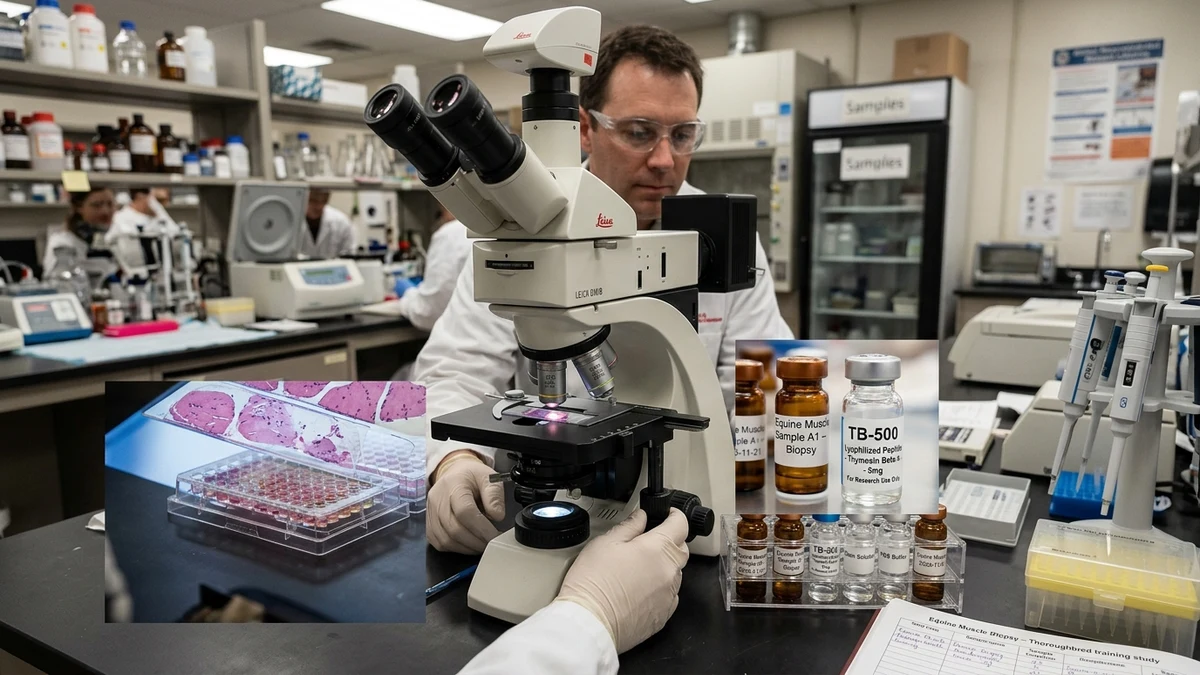
TB-500 occupies a specific and sometimes misunderstood position in the research landscape. It's a synthetic analog of the naturally occurring thymosin beta-4 peptide, typically produced via solid-phase peptide synthesis to a research grade. It is not approved as a veterinary therapeutic in most jurisdictions. In equine sports medicine, it appears on prohibited substance lists maintained by organizations including the FEI (Fédération Equestre Internationale) and various national racing authorities, which reflects both its use in competitive horses and the regulatory principle that unapproved biological compounds require prohibition pending proper clinical evaluation.
For legitimate pre-clinical research, this regulatory context means that investigators working with TB-500 in equine or other large-animal models operate within institutional animal care and use frameworks, with appropriate sourcing, documentation, and ethical oversight. The research value of such work is real. Large-animal pre-clinical data is a necessary step between rodent findings and any eventual therapeutic development, and horses, given their shared tendon pathology with humans, represent a scientifically meaningful bridge species.
The gap between what rodent models show and what can be claimed for clinical veterinary practice remains wide. That's not a failure of the science. It's the normal state of a research field that's still building its evidentiary foundation. What the existing data does support is continued, well-designed investigation into how thymosin beta-4 interacts with equine musculoskeletal tissue, under controlled conditions, with rigorous outcome measures. The biology is coherent, the comparative anatomy is favorable, and the clinical need in horses is undeniable. Those three things together make this a research area worth watching carefully.
This article is for informational and research purposes only. Nothing in this content constitutes veterinary or medical advice, a treatment recommendation, or an endorsement of any specific compound, supplier, or protocol. Research peptides are investigational substances not approved for veterinary therapeutic use in most jurisdictions. Always consult a licensed veterinarian before making any clinical decisions. For research purposes only, not veterinary advice.