Epithalon animal aging research has accumulated a surprisingly detailed body of work over the past four decades, much of it originating from Soviet and Russian biogerontology programs that studied the peptide's effects on pineal gland function, telomere dynamics, and age-related physiological decline. The tetrapeptide itself, Ala-Glu-Asp-Gly, was first synthesized by Vladimir Khavinson and colleagues at the St. Petersburg Institute of Bioregulation and Gerontology, and it remains one of the more studied short peptides in preclinical longevity science. What makes this body of literature worth examining is the consistency of certain findings across different animal models, though the research carries significant limitations that deserve honest acknowledgment.
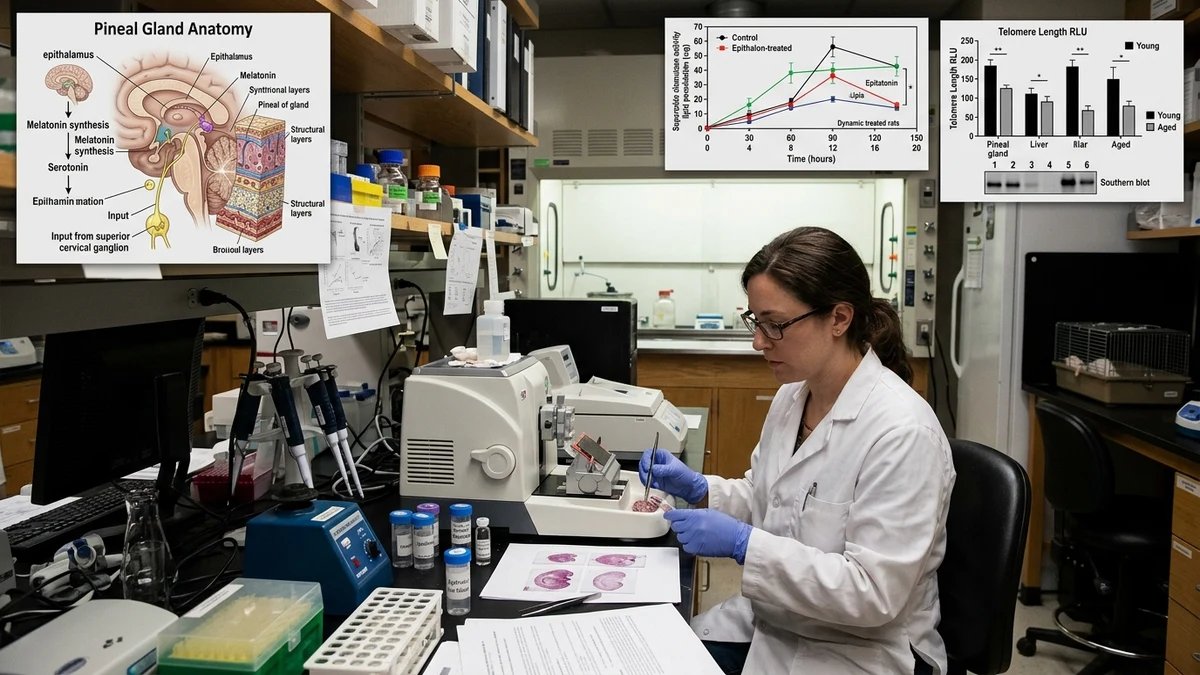
The pineal gland's role in biological aging has been a subject of scientific interest since at least the 1950s. The gland produces melatonin, regulates circadian rhythms, and appears to coordinate a broader orchestra of neuroendocrine signaling that shifts substantially as animals age. In rodents, pineal calcification and reduced melatonin output correlate with accelerated aging phenotypes. Epithalon was originally derived conceptually from the pineal gland's own peptide fractions, and early animal research focused on whether administering it exogenously could partially restore pineal-related signaling in aged subjects.
Studies conducted in aged rats and mice showed that epithalon administration was associated with increased melatonin synthesis, at least in animals whose baseline melatonin production had declined with age. The proposed mechanism involves epithalon's ability to influence the activity of hydroxyindole-O-methyltransferase, the enzyme responsible for the final step in melatonin biosynthesis. Research suggests this effect is not uniform across age groups, appearing more pronounced in subjects with already-suppressed pineal activity rather than in younger animals with functional glands.
Khavinson's group also documented changes in circadian rhythm regularity in treated aged rodents, with some subjects showing patterns of locomotor activity and sleep-wake cycling that more closely resembled younger animals. These findings are interesting, though they come primarily from a single research institution, and independent replication outside Russian institutions has been limited. That's not a reason to dismiss the data outright, but it's a genuine methodological limitation the field acknowledges.
Perhaps the most cited dimension of epithalon animal aging research is its apparent relationship with telomere biology. Telomeres, the protective caps on chromosomal ends, shorten with each cell division and serve as a rough biological clock for cellular aging. When telomeres erode past a critical threshold, cells enter senescence or apoptosis. Telomerase, the enzyme that can lengthen telomeres, is generally suppressed in somatic cells but active in germline cells and certain stem cell populations.
Research published by Khavinson and collaborators described epithalon's capacity to activate telomerase in somatic cell lines in vitro, with some follow-up observations in animal tissue. The proposed mechanism is indirect: epithalon appears to influence chromatin remodeling, potentially making the telomerase gene more accessible for transcription. This is a plausible biological pathway, and it connects to a broader area of epigenetic research on how short peptides might alter gene expression without acting as direct ligands to nuclear receptors.
In animal aging models, older mice and rats treated with epithalon showed tissue samples where telomere length was somewhat preserved compared to age-matched controls. The key word is "somewhat." The differences observed were statistically meaningful in several published papers, but the absolute magnitude was modest, and translating telomere preservation in rodent tissue to functional longevity outcomes requires careful interpretation. Rodents and humans have markedly different baseline telomere lengths and different telomerase regulation, which is one reason caution is warranted before drawing broad conclusions.
This connects naturally to related research areas like BPC-157 tissue repair studies and thymosin peptide research, where investigators are similarly trying to understand how short peptide sequences might influence cellular housekeeping mechanisms at the level of gene expression rather than direct receptor binding.
Some of the most provocative findings in epithalon animal aging research come from long-term rodent lifespan studies. Khavinson's group reported that both mice and rats receiving periodic epithalon injections over their natural lifespans showed modest but consistent increases in mean and maximum lifespan compared to untreated controls. The reported increases varied across individual studies, but research suggests the pattern was reproducible within that laboratory's experimental conditions.
Beyond raw lifespan, researchers tracked tumor incidence as a secondary endpoint. Rodents are prone to spontaneous tumor development, particularly mammary and liver tumors in certain strains, and age-related immunosenescence is a major factor in this susceptibility. Treated animals showed lower rates of spontaneous tumor development in several published reports. The immunological component here is worth unpacking. Epithalon treatment appeared to correlate with better-preserved immune function in aged rodents, including more stable natural killer cell activity and reduced markers of chronic low-grade inflammation.
Whether this represents a direct immunomodulatory effect or a downstream consequence of improved pineal and circadian function is not resolved. Chronic circadian disruption is itself a known driver of immune dysfunction and elevated cancer risk in animal models, so it's plausible these phenomena are linked rather than parallel. This is one area where the mechanistic picture remains genuinely unclear.
It's also worth situating these findings alongside research on thymic peptides in aging models, since thymosin alpha-1 and related compounds have shown overlapping effects on age-related immune decline. Epithalon isn't operating in an isolated biological context; it likely interacts with a web of aging-related processes that researchers are still mapping.
A line of research that has gained traction more recently examines how epithalon might influence gene expression patterns in aging tissue. The initial hypothesis, based on early in vitro work, was that the peptide acts as a chromatin-remodeling agent, reducing the compaction of heterochromatin in a way that allows certain silenced genes to become transcriptionally active again. In aged cells, gene silencing often extends beyond senescent or damaged genes to affect genes associated with repair, autophagy, and mitochondrial function.
Animal studies measuring messenger RNA expression in aged rodent tissue after epithalon treatment have shown differential expression of genes involved in oxidative stress response, DNA repair pathway activity, and antioxidant enzyme production. Superoxide dismutase and catalase activity specifically appeared elevated in treated aged animals relative to controls in some published work. These are real and measurable endpoints, which gives the epigenetic hypothesis more traction than pure speculation, though the studies were not large and the methods were not always fully described.
This epigenetic angle also connects to broader questions in peptide bioregulation research. Khavinson's theoretical framework, which he termed "peptide bioregulation," proposes that short peptides derived from specific tissues serve as natural modulators of gene expression in those tissues, and that declining peptide availability with age contributes to the disordered gene expression that characterizes aging. Whether this framework is ultimately validated or partially revised, it provides a coherent hypothesis for why a four-amino-acid sequence might show the kinds of effects reported in animal models.
The most honest assessment of epithalon animal aging research acknowledges a central limitation: the overwhelming majority of published studies come from one research group or its direct collaborators. This isn't unique to epithalon; many bioregulatory peptides have a similar publication history. But it does mean the dataset lacks the independent replication that would allow higher confidence in the findings.
Animal model selection also matters. Much of the foundational work used inbred rat and mouse strains that age in specific, somewhat predictable ways. These models are practical for research, but they don't always predict outcomes in outbred animals or humans with more heterogeneous genetic backgrounds. The jump from an aging Sprague-Dawley rat to a human with decades of accumulated environmental exposures and comorbidities is substantial.
Dosing regimens in animal studies are also difficult to translate directly. Researchers typically use weight-based dosing in rodents, and interspecies scaling for peptides is not straightforward because differences in renal clearance, receptor density, and metabolic rate all affect how much peptide actually reaches target tissues and for how long.
One concrete area where the research would benefit from expansion is in primate models. A small number of studies have explored epithalon in non-human primates, with some encouraging preliminary findings, but the sample sizes are too small and the follow-up durations too short to draw firm conclusions. Primate aging models would offer a much closer analog to human physiology and could clarify whether the pineal and telomere effects observed in rodents are genuinely conserved across mammalian species.
Researchers interested in peptide aging science will also find overlapping questions in IGF-1 pathway modulation studies and in literature on caloric restriction mimetics, since many of the cellular endpoints, telomere preservation, reduced oxidative stress, maintained immune function, appear across multiple longevity-focused research areas. Epithalon's proposed mechanisms are not biologically isolated; they fit within a larger framework of interventions thought to slow cellular aging processes.
Preclinical evidence for epithalon's effects on pineal function, telomere biology, and aging-related gene expression in animal models is more developed than most people in the general research community realize. The work is not preliminary in the sense of a handful of exploratory studies; it represents decades of systematic investigation with consistent directional findings across several biological endpoints.
At the same time, the field carries real scientific debt in the form of limited independent replication and an over-reliance on a narrow set of animal models. The pineal-aging hypothesis that underlies much of this research is plausible and well-grounded in circadian biology, but the pathway from pineal restoration to whole-organism longevity involves enough intermediate steps that simple causal stories are almost certainly incomplete.
What the animal aging literature on epithalon does offer is a foundation for more rigorous inquiry. The biological endpoints are measurable, the proposed mechanisms are falsifiable, and the research tradition is long enough to identify which findings have held up across different experimental conditions. That's more than can be said for many compounds with much larger public profiles.
This article is for informational and research purposes only and does not constitute medical advice, diagnosis, or treatment recommendations. Epithalon is a research compound studied in preclinical and animal models. Any research involving peptides should be conducted under appropriate institutional and regulatory oversight. For research purposes only — not medical advice.