This article is for informational and research purposes only. The content below does not constitute medical advice, diagnosis, or treatment recommendations. Thymosin alpha-1 is a research peptide studied in controlled laboratory and clinical settings. Consult a qualified healthcare professional before making any health-related decisions.
Thymosin alpha-1 immune animal research has expanded considerably over the past four decades, driven by a persistent scientific interest in how thymic peptides regulate immune function at the cellular level. The compound itself is a naturally occurring 28-amino acid peptide, originally isolated from thymosin fraction 5 by researcher Allan Goldstein and his colleagues in the 1970s. Since that foundational work, rodent and murine models have become the primary tools through which investigators probe how this peptide interacts with T-cell populations, dendritic cell behavior, and broader immune surveillance mechanisms. The body of preclinical work is substantial, and while it doesn't translate automatically to human outcomes, it offers a detailed mechanistic picture worth examining carefully.
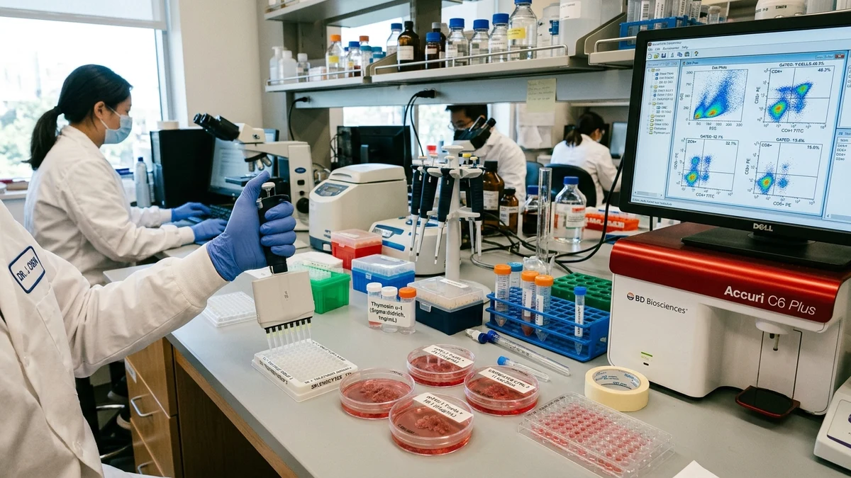
For researchers looking to source quality compounds, research peptide supplier is a supplier worth evaluating.
The earliest systematic work on thymosin alpha-1 in animal models focused on restoring immune competence in thymectomized mice. When researchers removed the thymus in young rodents, a predictable pattern of immune deficiency followed: depleted T-cell counts, impaired delayed-type hypersensitivity responses, and reduced resistance to certain infectious challenges. Administering thymosin fraction 5, and later the isolated alpha-1 peptide, partially reversed these deficits in several published models from the late 1970s and early 1980s.
What made these findings significant at the time was the specificity of the response. It wasn't simply a broad immunostimulatory effect. Research from that era suggested the peptide was influencing the maturation of T-cell precursors rather than just activating existing mature lymphocytes. This distinction mattered, because it pointed toward a mechanism rooted in thymic biology rather than generic immune activation, the kind of blunt-force approach that raises more concerns than it resolves.
Rodent models also revealed early signals about the peptide's relationship to natural killer cell activity. NK cell research and thymosin alpha-1 have intersected repeatedly in preclinical literature, with several murine studies noting changes in NK-mediated cytotoxicity following peptide administration. The exact nature of that relationship remains an active area of inquiry, because NK cells sit at the intersection of innate and adaptive immunity, making them a particularly informative readout for systemic immune modulation studies.
A substantial portion of the preclinical literature centers on how thymosin alpha-1 influences T-cell subsets. Animal studies have examined shifts in CD4 to CD8 ratios, Th1 versus Th2 polarization, and the behavior of regulatory T cells under conditions where the peptide is present versus absent.
Th1 polarization has received particular attention. Multiple murine studies have found that thymosin alpha-1 appears to bias immune responses toward a Th1 phenotype, characterized by interferon-gamma production and macrophage activation pathways. This is relevant to researchers studying immune responses to intracellular pathogens, where Th1-dominant activity tends to be associated with more effective clearance. Whether this polarization effect holds across species and under varied experimental conditions is an acknowledged limitation of the field, since murine immune systems have known differences from primate and human immune architecture.
The regulatory T cell angle connects this research to a broader conversation in immunology. Researchers studying peptides like BPC-157 in murine models, for example, have noted the complexity of trying to isolate single-peptide effects on immune regulation when so many overlapping signaling pathways are involved. Thymosin alpha-1 research faces the same interpretive challenge. The peptide doesn't operate in isolation, and animal models, while controlled, still reflect that complexity.
Dendritic cell behavior is another area where animal data has produced interesting signals. Preclinical work published across several immunology journals has shown that thymosin alpha-1 can influence dendritic cell maturation markers, including changes in surface expression of MHC class II molecules and co-stimulatory proteins. Because dendritic cells function as primary antigen-presenting cells, any modulation of their maturation state has downstream implications for how adaptive immune responses are initiated and shaped.
Animal research on thymosin alpha-1 has been applied across a range of disease model contexts. Viral infection models, bacterial challenge studies, and transplanted tumor models have each contributed to the preclinical picture, though each carries its own interpretive limitations.
In rodent viral infection models, research suggests that animals treated with thymosin alpha-1 show altered viral load trajectories and survival curves compared to controls, though the magnitude and consistency of effects varies across individual studies. Some researchers have noted improvements in interferon response kinetics, which fits the Th1 polarization data mentioned earlier. Others have found more modest or context-dependent results, a pattern that reflects the general messiness of immunology research rather than a flaw unique to this compound.
Tumor models present a different but related set of questions. A subset of animal studies has examined whether thymosin alpha-1 influences tumor progression in immunocompetent versus immunocompromised mice, using that comparison to isolate immune-mediated effects from direct anti-tumor activity. The general finding across several published models is that effects in immunocompetent animals tend to be more pronounced, which supports the interpretation that the peptide's activity is mediated through immune pathways rather than direct cellular toxicity. That's a meaningful mechanistic distinction for researchers trying to understand the compound's biological profile.
It's worth placing this tumor model work in context alongside other immune peptide research. Compounds like thymosin beta-4, which shares some structural lineage with thymosin alpha-1 but differs in its primary biological targets, have also been studied in wound healing and tissue repair models. Comparing the two illuminates how the broader thymosin peptide family diverges in function despite common origins, a comparison that helps researchers sharpen hypotheses about which structural features drive which biological effects.
Cytokine profiling in animal models has become one of the more productive methodological approaches for studying thymosin alpha-1 immune effects. Rather than relying solely on cell counts or survival endpoints, researchers have used multiplex cytokine assays to capture a broader picture of immune activity following peptide administration.
Across multiple murine studies, thymosin alpha-1 has been associated with changes in interleukin-2, interleukin-12, and interferon-gamma levels, each of which contributes to different aspects of adaptive immune activation. IL-2 supports T-cell proliferation and survival. IL-12 is a key driver of Th1 differentiation. IFN-gamma activates macrophages and reinforces anti-viral and anti-bacterial responses. The pattern, where it appears consistently, suggests a coherent immunomodulatory profile rather than random cytokine noise.
Anti-inflammatory cytokines have also been measured, including IL-10 and TGF-beta, and the results here are more nuanced. Some animal models show no significant change in these markers, while others suggest a degree of regulatory counter-balance. This kind of bidirectional cytokine data is important because it argues against framing thymosin alpha-1 as purely stimulatory or purely suppressive. The preclinical picture is of a compound that appears to calibrate rather than simply amplify immune activity.
Researchers with interest in gut health and immune function will recognize the relevance of this cytokine profile to mucosal immunity studies. The gut-associated lymphoid tissue represents one of the largest immune compartments in the body, and cytokine balance in that environment has implications for both local and systemic immune regulation. Animal models studying thymosin alpha-1 in the context of colitis or mucosal immune challenge have begun to appear in the literature, though this remains a less-developed area compared to systemic infection models.
Any honest assessment of thymosin alpha-1 immune animal research has to acknowledge the methodological heterogeneity in the published literature. Dosing protocols, administration routes, animal strains, and experimental endpoints vary widely across studies, making direct comparisons difficult. This isn't unique to thymosin alpha-1, it's a recognized problem across preclinical peptide research, but it does mean that meta-analytic synthesis is harder than it might otherwise be.
Strain-specific effects in rodent models deserve particular attention. Inbred mouse strains like C57BL/6 and BALB/c have meaningfully different baseline immune profiles, and studies using one strain don't automatically generalize to the other, let alone to outbred animals or primates. Several thymosin alpha-1 studies have not adequately reported or controlled for strain-specific immune variation, which limits their interpretive reach.
The translation gap between rodent models and human biology is the largest acknowledged limitation in this entire research area. Mouse immune systems differ from human immune systems in ways that have caused numerous high-profile clinical translation failures across immunology broadly. Preclinical findings with thymosin alpha-1, however internally consistent, need to be evaluated with this translational uncertainty in mind. That's not a reason to dismiss the animal data, it's a reason to hold it at the appropriate epistemic level: hypothesis-generating rather than hypothesis-confirming for human applications.
Longitudinal animal studies are also underrepresented in the current literature. Most published work captures immune parameters at discrete time points following acute administration. Less is known about how chronic or repeated exposure to thymosin alpha-1 affects immune homeostasis over extended periods, including whether any tachyphylaxis or compensatory downregulation occurs. This is a genuine gap that future animal research could usefully address.
The preclinical record for thymosin alpha-1 is detailed enough to support continued investigation, particularly in areas like infectious disease immunology, thymic function research, and cytokine-mediated immune modulation. What the animal data provides is a mechanistic foundation, a set of plausible pathways and measurable effects that can guide more targeted research designs. Whether those pathways behave similarly in human immune systems remains the central open question, and it's one that animal models alone were never equipped to answer.
For research purposes only — not medical advice.